-
Find the right food for your petTake this quiz to see which food may be the best for your furry friend.Find the right food for your petTake this quiz to see which food may be the best for your furry friend.Health CategoryFeatured products
 Adult Salmon & Brown Rice Recipe Dog Food
Supports lean muscle and beautiful coat for adult dogs
Shop Now Adult Sensitive Stomach & Skin Chicken Recipe Dog Food
Hill's Science Diet Sensitive Stomach & Skin dry dog food is gentle on stomachs while nourishing skin & promoting a lustrous coat.
Shop Now Adult Healthy Mobility Chicken Meal, Barley & Brown Rice Recipe Dog Food
Advanced nutrition shown to support joint health and improve mobility
Shop NowFeatured products Adult Salmon & Brown Rice Recipe Cat Food
Supports lean muscle and beautiful fur for adult cats
Shop Now Adult Perfect Weight with Chicken Cat Food
Breakthrough nutrition for your cat’s healthy weight maintenance and long-lasting weight support
Shop Now Adult Urinary Hairball Control Tender Chicken Dinner Cat Food
Precisely balanced nutrition to support urinary health from kidney to bladder. With natural fibre technology to help reduce hairballs.
Shop Now -
DogCat
- Cat Tips & Articles
-
Health Category
- Weight
- Skin & Food Sensitivities
- Urinary
- Digestive
- Kidney
- Dental
- Serious Illness
-
Life Stage
- Kitten Nutrition
- Adult Nutrition
Featured articles The Right Diet For Your Pet
Learn what to look for in healthy pet food & nutrition, including ingredients, quality of the manufacturer, your pet's age, and any special needs they have.
Read More Pet Food Storage Tips
Where you store your cat and dog food can make a big difference in the quality and freshness once it is opened. Here are some common questions and recommendations for optimal storage for all of Hill’s dry and canned cat and dog food.
Read More Water
WaterWater is the most important nutrient of all and essential for life. Animals can lose almost all their fat and half their protein and still survive, but if they lose 15% of their water, it will mean death.
Read More -


Mast cell tumors in dogs are the No. 1 most common skin tumor in dogs, according to the American College of Veterinary Surgeons. But what exactly is a mast cell tumor, what are the signs of mast cell tumors in dogs and how are these tumors treated?
Causes of Mast Cell Tumors in Dogs

Mast cells themselves aren't cancer cells. As the National Cancer Institute explains, they're a type of white blood cell, though they aren't normally found in the blood. They originate in the bone marrow and are found throughout the body's various tissues. Mast cells are normally involved in a number of functions in your dog. They play a role in inflammation, allergic responses, parasite presence responses, and even necrosis and the breakdown of dead tissues.
Unfortunately, sometimes things go awry. For reasons veterinary scientists don't yet understand, mast cells sometimes mutate and multiply in masses, going against the normal cell life cycle that keeps their numbers in check. This unregulated growth results in a mast cell tumor. These tumors can occur in dogs of all breeds and ages.
Clinical Signs of Mast Cell Tumor in Dogs
Mast cell tumors in dogs can vary in appearance, but they're usually in the form of a lump. These lumps can occur on the skin, muzzle, mouth, genitals or even inside the body on the organs.
When you're petting or examining your dog, you may notice a firm lump tightly adhered to the skin or a squishy and movable lump under the skin. You may see one or multiple lumps, and they might be ulcerated, oozing or bleeding. The lump may remain the same size, grow rapidly or even recede, but that doesn't make it any less dangerous.


Tasty Tips
Diagnosing Mast Cell Tumors in Dogs
Lumpy, bumpy, raised, flat, loose, squishy, firm, big or small — each and every lump on your dog should be evaluated by a veterinarian.
Your vet will need to perform a diagnostic test called a fine needle aspiration to collect a small sampling of cells from the tumor. Luckily, this test is fast and minimally invasive, and it can usually be done following the initial examination. However, some tumors don't shed many cells during this test, or the sample may not be representative of the whole lesion. So, though this test is a great starting point, your vet may recommend a punch biopsy, as it allows a more comprehensive examination of the mass's architecture. With a biopsy, a microscopic examination of the tissue (histopathology) is sent out to a specialized laboratory for a veterinary pathologist to prepare and evaluate.
After a mast cell tumor diagnosis, your vet may recommend additional diagnostics such as bloodwork, chest radiographs (X-rays) and an abdominal ultrasound to determine whether the tumor has spread to other locations (known as metastasis). A fine needle aspirate of a local lymph node may also be recommended.

Mast Cell Tumor Treatment
To determine the best treatment for a mast cell tumor, an expert reading from a board-certified veterinary pathologist is necessary. All recommendations for cancer treatment in dogs require a pathologist to determine the stage, or extent, of the cancer. Mast cell tumors are graded from I to III, with I being the least aggressive and III being the most advanced. The other diagnostics (X-rays, lymph node aspiration, bloodwork and imaging as needed) allow the pathologist to formulate a top-notch treatment plan.
Depending upon the report, the pathologist may recommend surgery, with or without chemotherapy. After surgery, the mass should be sent in for a report again to ensure that all the cancerous cells were removed and clean margins (meaning there aren't cancer cells at the outer edge of the tissue) were obtained, which isn't always possible. Note that while surgery and chemotherapy are often recommended for tumors in grades II and III, you can also discuss less aggressive approaches aimed more at comfort care at any stage with your vet and/or veterinary oncologist.
Life Expectancy for Dogs With Mast Cell Tumors
It is difficult to predict a single life expectancy for dogs who develop mast cell tumors, as it varies depending on many factors. If the tumor is limited to the skin with no evidence of metastasis and surgical removal achieved clean margins, the prognosis can be quite good. For tumors that are scored a grade II or III, the prognosis may not be as good. Furthermore, tumors in locations such as the mouth or genitals often lead to a more guarded prognosis.
The good news is that early diagnosis and treatment improve the prognosis of mast cell tumors, making it important to regularly examine your dog at home. This can be done simply with observational petting and intentional at-home exams. Remember to schedule a veterinary visit should you discover any new masses on your dog.


Dr. Laci Schaible is a small animal veterinarian, veterinary journalist, and a thought leader in the industry. She received her Doctor of Veterinary Medicine from Texas A&M University and her Masters in Legal Studies from Wake Forest University.
Related products

Gentle on stomachs while nourishing skin & supporting development in growing puppies

Supports lean muscle and beautiful coat for adult dogs

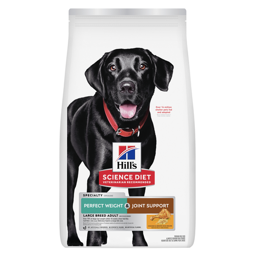
This weight management and mobility support dog food was created with Hill’s unique understanding of the biology of overweight dogs

Delicious roasted chicken paired with tender vegetables in a succulent stew
Related articles

Though it may seem like your four-legged friend loves nothing more than to nap on the couch, dogs need regular exercise to stay healthy just like people do.

A dog with a sensitive stomach has special needs. Learn more about sensitive stomach symptoms in your dog, what you can do to help sooth your pet’s insides and get recommendations on sensitive stomach dog food.

Selecting the right food for your puppy is a key to quality nutrition and a long, healthy life., Learn more about how to select the right puppy food.

Learn what you can feed your pregnant or nursing dog to keep her and her new pups healthy.

Put your dog on a diet without them knowing
Our low calorie formula helps you control your dog's weight. It's packed with high-quality protein for building lean muscles, and made with purposeful ingredients for a flavorful, nutritious meal. Clinically proven antioxidants, Vitamin C+E, help promote a healthy immune system.

